39 microscope diagram labelled
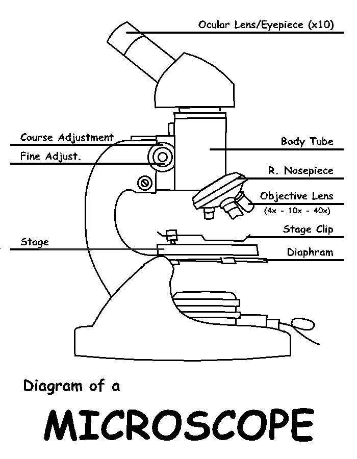
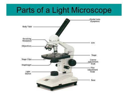
Microscope Parts and Functions It also allows the specimen to be labeled, transported, and stored without damage. Stage: The flat platform where the slide is placed. Stage clips: Metal clips that hold the slide in place. Stage height adjustment (Stage Control): These knobs move the stage left and right or up and down. PDF Label parts of the Microscope: Answers Label parts of the Microscope: Answers Coarse Focus Fine Focus Eyepiece Arm Rack Stop Stage Clip . Created Date: 20150715115425Z ...
Microscope Types (with labeled diagrams) and Functions Simple microscope labeled diagram Simple microscope functions It is used in industrial applications like: Watchmakers to assemble watches Cloth industry to count the number of threads or fibers in a cloth Jewelers to examine the finer parts of jewelry Miniature artists to examine and build their work Also used to inspect finer details on products
Microscope diagram labelled
Simple Microscope - Parts, Functions, Diagram and Labelling Parts of the optical parts are as follows: Mirror - A simple microscope has a plano-convex mirror and its primary function is to focus the surrounding light on the object being examined. Lens - The biconvex lens is placed above the stage and its function is to magnify the size of the object being examined. Light microscopes - Cell structure - Edexcel - BBC Bitesize Microscopes use lenses to magnify the image of a specimen so that it appears larger. The formula to calculate magnification is: \ [\text {magnification} = \frac {\text {size of image}} {\text {real... Microscope Diagram Labeled, Unlabeled and Blank | Parts of a Microscope ... Worksheet identifying the parts of the compound light microscope. Answer key: 1. Body tube 2. Revolving nosepiece 3. Low power objective 4. Medium power objective 5. High power objective 6. Stage clips 7. Diaphragm 8. Light source 9. Eyepiece 10. Arm 11. Stage 12. Coarse adjustment knob 13. Fine ad... S Stephanie Coville Microscope Biology Test
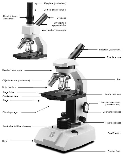
Microscope diagram labelled. Label the microscope — Science Learning Hub Use this interactive to identify and label the main parts of a microscope. Drag and drop the text labels onto the microscope diagram. base stage high-power objective eye piece lens coarse focus adjustment light source diaphragm or iris fine focus adjustment Download Exercise Tweet Compound Microscope Parts, Functions, and Labeled Diagram Compound Microscope Definitions for Labels Eyepiece (ocular lens) with or without Pointer: The part that is looked through at the top of the compound microscope. Eyepieces typically have a magnification between 5x & 30x. Monocular or Binocular Head: Structural support that holds & connects the eyepieces to the objective lenses. Compound Microscope Parts - Labeled Diagram and their Functions Labeled diagram of a compound microscope Major structural parts of a compound microscope There are three major structural parts of a compound microscope. The head includes the upper part of the microscope, which houses the most critical optical components, and the eyepiece tube of the microscope. Parts of Stereo Microscope (Dissecting microscope) - labeled diagram ... Labeled part diagram of a stereo microscope Major structural parts of a stereo microscope There are three major structural parts of a stereo microscope. The viewing Head includes the upper part of the microscope, which houses the most critical optical components, including the eyepiece, objective lens, and light source of the microscope.
Microscope Diagram - cell division of e coli with continuous media flow ... Microscope Diagram - 15 images - give a well labelled diagram of compound microscope using of typical, bio tem biological transmission electron microscope university, labelled microscope diagram gcse micropedia, a compound microscope diagram micropedia, Labelled Diagram of Compound Microscope - Biology Discussion The below mentioned article provides a labelled diagram of compound microscope. Part # 1. The Stand: The stand is made up of a heavy foot which carries a curved inclinable limb or arm bearing the body tube. The foot is generally horse shoe-shaped structure (Fig. 2) which rests on table top or any other surface on which the microscope in kept. (a) Draw the labelled ray diagram for the formation of image by a ... Click here👆to get an answer to your question ️ (a) Draw the labelled ray diagram for the formation of image by a compound microscope. Derive an expression for its total magnification (or magnifying power), when the final image is formed at the near point.(b) Why both objective and eyepiece of a compound microscope must have short focal lengths?Draw a ray diagram showing the image ... Parts of a Compound Microscope and Their Functions - NotesHippo Compound microscope mechanical parts (Microscope Diagram: 2) include base or foot, pillar, arm, inclination joint, stage, clips, diaphragm, body tube, nose piece, coarse adjustment knob and fine adjustment knob. Base: It's the horseshoe-shaped base structure of microscope. All of the other components of the compound microscope are supported by it.
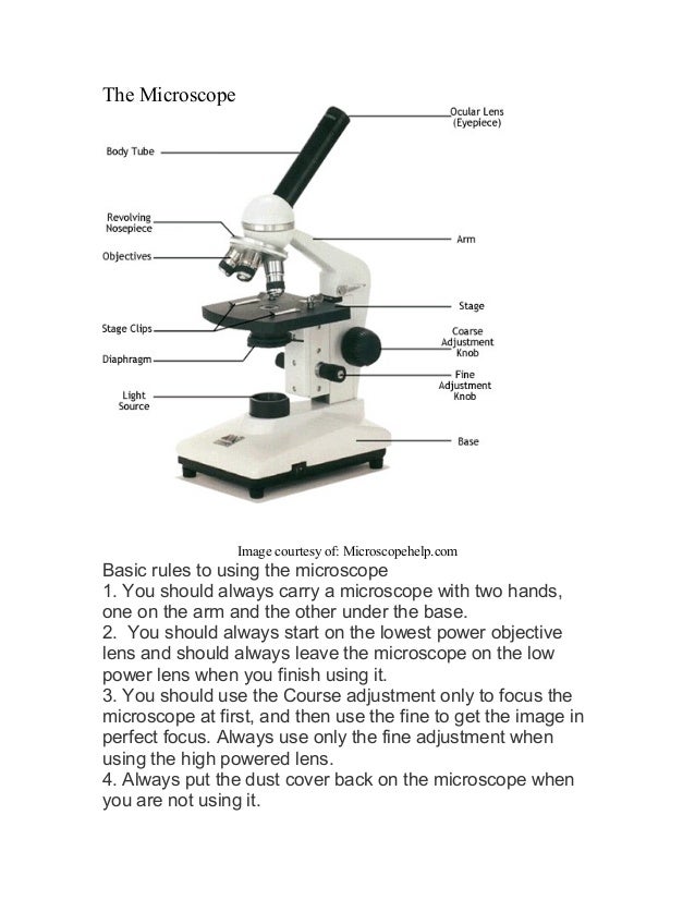
Microscope labeled diagram - SlideShare 1. The Microscope Image courtesy of: Microscopehelp.com Basic rules to using the microscope 1. You should always carry a microscope with two hands, one on the arm and the other under the base. 2. You should always start on the lowest power objective lens and should always leave the microscope on the low power lens when you finish using it. 3. Microscope, Microscope Parts, Labeled Diagram, and Functions Microscope, Microscope Parts, Labeled Diagram, and Functions What is Microscope? A microscope is a laboratory instrument used to examine objects that are too small to be seen by the naked eye. It is derived from Ancient Greek words and composed of mikrós, "small" and skopeîn,"to look" or "see". Neuron under Microscope with Labeled Diagram - AnatomyLearner Neuron under microscope labelled diagram. Throughout this article, you got the different neurons labelled diagrams. Here, you will also find the diagrams of different neuron types under a microscope. The neuron diagram shows the different parts (axon, dendrites, and cell body) of the neurons. The Parts of a Microscope (Labeled) Printable - TeacherVision The Parts of a Microscope (Labeled) Printable. Download. Add to Favorites. Share. This diagram labels and explains the function of each part of a microscope. Use this printable as a handout or transparency to help prepare students for working with laboratory equipment.
Diagram of a Microscope by ScienceDoodles on DeviantArt
Compound Microscope- Definition, Labeled Diagram, Principle, Parts, Uses There are two basic types of optical microscopes: Simple microscopes. Compound microscopes. The term "compound" in compound microscopes refers to the microscope having more than one lens. Devised with a system of combination of lenses, a compound microscope consists of two optical parts, namely the objective lens and the ocular lens.

Label the image of a compound light microscope - AnswerPrime.com
Microscope Labeling Diagram | Quizlet Unit 2 Lesson 5 - Punnett Squares and Pedigrees. 4 terms. PGFry210. Unit 2 Lesson 4 - Heredity. 9 terms. PGFry210. Upgrade to remove ads. Only $2.99/month.

Parts of a Compound Microscope and Their Functions
Microscope Parts, Function, & Labeled Diagram - slidingmotion Microscope parts labeled diagram gives us all the information about its parts and their position in the microscope. Microscope Parts Labeled Diagram The principle of the Microscope gives you an exact reason to use it. It works on the 3 principles. Magnification Resolving Power Numerical Aperture. Parts of Microscope Head Base Arm Eyepiece Lens

Parts of the Microscope with Labeling (also Free Printouts ...
Microscopes - Cell structure - AQA - GCSE Combined Science ... - BBC Late 1600s - Dutch scientist Antonie van Leeuwenhoek constructed a microscope with a single spherical lens. It magnified up to ×275. 1800s - the optical quality of lenses increased and the ...

Microscope Diagram – Charts
Parts of a microscope with functions and labeled diagram - Microbe Notes Figure: Diagram of parts of a microscope There are three structural parts of the microscope i.e. head, base, and arm. Head - This is also known as the body. It carries the optical parts in the upper part of the microscope. Base - It acts as microscopes support. It also carries microscopic illuminators.

5 Important Types of Microscopes used in Biology (With Diagram)
PDF Basic Histo diagrams labelled in colour - 2005 - UNSW Sites These labelled diagrams should closely follow the current Science courses in histology, anatomy and ... Do microscope images of 2-D slices represent a single plane of section of a 3-D structure? No, 2-D slices have a thickness which can vary from a sliver of one cell to several cells deep.

This is a common compound microscope. Label its parts from A ...
A Study of the Microscope and its Functions With a Labeled Diagram ... A Study of the Microscope and its Functions With a Labeled Diagram To better understand the structure and function of a microscope, we need to take a look at the labeled microscope diagrams of the compound and electron microscope. These diagrams clearly explain the functioning of the microscopes along with their respective parts.

Microscope Labeling
Simple Microscope - Diagram (Parts labelled), Principle, Formula and Uses Labeled Diagram of simple microscope parts Optical parts. The optical parts of a simple microscope include. Lens; Mirror; Eyepiece; Lens. A simple microscope uses biconvex lens to magnify the image of a specimen under focus.

Parts of a Microscope Labeling Activity
Parts of the Microscope with Labeling (also Free Printouts) Parts of the Microscope with Labeling (also Free Printouts) By Editorial Team March 7, 2022 A microscope is one of the invaluable tools in the laboratory setting. It is used to observe things that cannot be seen by the naked eye. Table of Contents 1. Eyepiece 2. Body tube/Head 3. Turret/Nose piece 4. Objective lenses 5. Knobs (fine and coarse) 6.
Parts of a Microscope and Their Functions
Labeling the Parts of the Microscope | Microscope World Resources Labeling the Parts of the Microscope This activity has been designed for use in homes and schools. Each microscope layout (both blank and the version with answers) are available as PDF downloads. You can view a more in-depth review of each part of the microscope here. Download the Label the Parts of the Microscope PDF printable version here.

Parts of a microscope with functions and labeled diagram
Microscope Diagram Labeled, Unlabeled and Blank - Pinterest timvandevall.com Microscope Diagram Labeled, Unlabeled and Blank | Parts of a Microscope - Tim's Printables Print a microscope diagram, microscope worksheet, or practice microscope quiz in order to learn all the parts of a microscope. T Tim's Printables 37k followers More information Microscope Diagram

Microscope labeling
Microscope Diagram Labeled, Unlabeled and Blank | Parts of a Microscope ... Worksheet identifying the parts of the compound light microscope. Answer key: 1. Body tube 2. Revolving nosepiece 3. Low power objective 4. Medium power objective 5. High power objective 6. Stage clips 7. Diaphragm 8. Light source 9. Eyepiece 10. Arm 11. Stage 12. Coarse adjustment knob 13. Fine ad... S Stephanie Coville Microscope Biology Test

Simple Microscope - Diagram (Parts labelled), Principle ...
Light microscopes - Cell structure - Edexcel - BBC Bitesize Microscopes use lenses to magnify the image of a specimen so that it appears larger. The formula to calculate magnification is: \ [\text {magnification} = \frac {\text {size of image}} {\text {real...

MICROSCOPE Drawing
Simple Microscope - Parts, Functions, Diagram and Labelling Parts of the optical parts are as follows: Mirror - A simple microscope has a plano-convex mirror and its primary function is to focus the surrounding light on the object being examined. Lens - The biconvex lens is placed above the stage and its function is to magnify the size of the object being examined.

Microscope Diagram Labeled, Unlabeled and Blank | Parts of a ...

Microscope With Labels Clip Art at Clker.com - vector clip ...

Light Microscope- Definition, Principle, Types, Parts ...

Labeled microscope diagram
Microscope Maintenance Tips
Anatomy of a Microscope | Microscopy Primer | Olympus LS

Microscope Isometric Illustration With Light Microscope Parts ...
Microscope labeled diagram
Microscope, Microscope Parts, Labeled Diagram, and Functions

draw a well label diagram of microscope - Brainly.in

The Microscope- compound microscope diagram - Major Science ...

Draw a neat labelled diagram of a compound microscope class ...

Produk Microscope | UD Berkah Abadi
Microscope Parts, Structure Anatomy. Vector 3d Realistic ...

Label Microscope Diagram - EnchantedLearning.com
Microscope - Teaching resources

Compound Microscope Parts, Diagram Definition, Application ...

Draw a labelled diagram of a compound microscope.

Microscope Diagram Labeled Parts - ClipArt Best - ClipArt ...

Microscope Parts Diagram PDF – Tim's Printables

How to Use a Microscope: Learn at Home with HST Learning ...
Vektor Stok Microscope Diagram Vector Illustration Labeled ...
Calibration of Microscopes and Scale Drawings. - ppt video ...

The schematic of the combined optical and acoustic microscope ...
Microscopy- History, Classification, Terms, Diagram
Post a Comment for "39 microscope diagram labelled"