38 labelled femur
labeled femur Flashcards | Quizlet 18 terms Kimberly_Miller33 labeled femur STUDY PLAY Articular Cartilage Canaliculi Compact bone Diaphysis Distal diaphysis Endosteum Epiphyseal plate Haversian canal Lacuna Medullary cavity Osteocyte Osteon Periosteum Proximal epiphysis Spongy bone Red marrow is found here Volkmann's canal Yellow marrow is found here Features Quizlet Live Femur Bone: Definition, Diagrams, Location, Parts & Functions The femur starts to evolve between the 5th to 6th gestational week by the path of endochondral ossification (where a bone is composed using a cartilage-depended foundation). While several ossification places (points of bone development) emerge went through intrauterine life, the bone continues to advance through childhood and early boyhood.Ossification of the femur is finished between the 14th ...
femur | Definition, Function, Diagram, & Facts | Britannica femur, also called thighbone, upper bone of the leg or hind leg. The head forms a ball-and-socket joint with the hip (at the acetabulum), being held in place by a ligament ( ligamentum teres femoris) within the socket and by strong surrounding ligaments.

Labelled femur
labeled femur bone - anatomyclass99.z21.web.core.windows.net Femur_labeled anatomycorner.com. femur labeled bones humerus human anatomy skull unlabeled skeleton radius ulna disarticulated anatomycorner main. Femur-Right Anterior View . drawing bones skeleton anatomy bone femur sketch body references printable drawings line thigh human character flash casson anatomi minimal inspiration Femur bone anatomy: Proximal, distal and shaft | Kenhub The femur bone is the strongest and longest bone in the body, occupying the space of the lower limb, between the hip and knee joints. Femur anatomy is so unique that it makes the bone suitable for supporting the numerous muscular and ligamentous attachments within this region, in addition to maximally extending the limb during ambulation. Femur Labeled Diagram | Quizlet Start studying Femur Labeled. Learn vocabulary, terms, and more with flashcards, games, and other study tools.
Labelled femur. Labeled Skeletal System Diagram - Bodytomy Bone Structure of the Chest and Hip. The bones shown in the chest and hip region in the labeled human skeleton diagram are the ribs, vertebrae, pelvis, OS coxae, sacrum and coccyx. Total there are 12 pairs of ribs, as you can see in the diagram. The last pair of the ribs, which is at the bottom of the rib, are called floating ribs, as they are ... The Femur - Human Anatomy The Femur. F IG. 243- Upper extremity of right femur viewed from behind and above. (Thigh Bone) The femur (Figs. 244, 245), the longest and strongest bone in the skeleton, is almost perfectly cylindrical in the greater part of its extent. In the erect posture it is not vertical, being separated above from its fellow by a considerable interval ... femur bone anatomy not labeled knee pain tibia fibula head lateral muscle labeled schlatter osgood disease femur laser hamstrings patellar joint patella femoris chiropractor anatomy. 19 New Parts Of Femur Bone rycaaschiffman.blogspot.com. Click On A Region In The Picture To Color It In With The Selected Color. Femur length (obstetric ultrasound) | Radiology Reference Article ... Femur length together with biparietal diameter, head circumference , and abdominal circumference are computed to produce an estimate of fetal weight. In the second trimester, this may be extrapolated to an estimate of gestational age and an estimated due date (EDD). Femur length can be used to calculate fetal length, another predictor of fetal ...
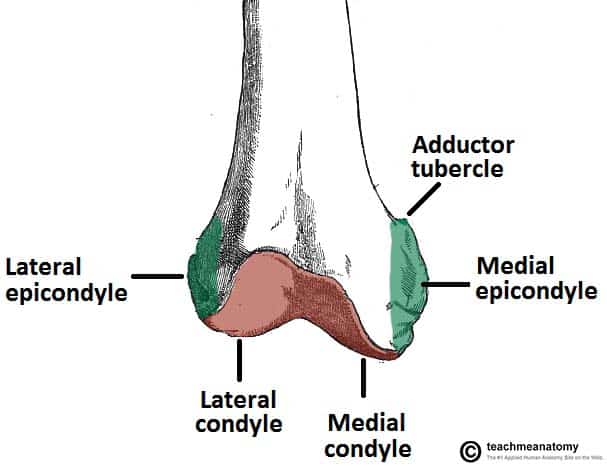
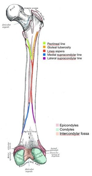
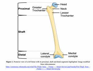
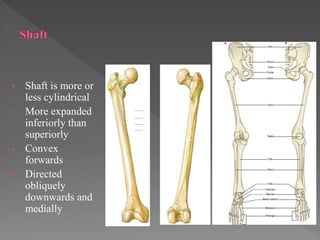
labelled femur bone labelled femur bone Femur Bone. 17 Images about Femur Bone : Femur Bone Cross Section - Cross Section Of The Head Of The Femur, Computational and experimental investigation on the effect of failure and also Frontal Section of Hip Joint | ClipArt ETC. Femur Bone › en › libraryLeg and knee anatomy: Bones, muscles, soft tissues | Kenhub Aug 02, 2022 · The main parts of the knee joint are the femur, tibia, patella, and supporting ligaments. The condyles of the femur and of the tibia come in close proximity to form the main structure of the joint. The patella, commonly known as the ‘kneecap’, is a sesamoid bone that sits within the tendon of the quadriceps femoris. It serves a protective ... Femur Bone Quiz - Posterior Markings | GetBodySmart Femur Bone Quiz - Posterior Markings . Start Quiz. Retake Quiz. Accelerate your understanding and retention with these interactive, exam-style anatomy quizzes. Learn anatomy faster and remember everything you learn. Start Now. Related Articles. Tibia and Fibula Quiz: Posterior Markings. Tibia and Fibula Bones Quiz - Anterior Markings ... The Femur - Proximal - Distal - Shaft - TeachMeAnatomy The femur is the only bone in the thigh and the longest bone in the body. It acts as the site of origin and attachment of many muscles and ligaments, and can be divided into three parts; proximal, shaft and distal. In this article, we shall look at the anatomy of the femur - its attachments, bony landmarks, and clinical correlations. Proximal
anatomylearner.com › horse-skeleton-anatomyHorse Skeleton Anatomy - Osteological Features of Bones from ... Nov 06, 2021 · The femur of horse anatomy. The femur is the largest and more massive bone in a horse skeleton. It extends obliquely distally and cranially. The femur articulates with the acetabulum proximally and the tibia and patella distally. You will find a cylindrical body and two large extremities in the femur of a horse. Learn femur anatomy fast with these femur quizzes | Kenhub The femur is a long bone found in the lower extremity. It serves as the attachment site for several muscles of the hip and leg, allowing it to withhold pressure from multiple angles. There are three main parts to the femur: The proximal end The shaft The distal end Femur (Thighbone): Anatomy, Function & Common Conditions - Cleveland Clinic Femur The femur is the longest, strongest bone in your body. It plays an important role in how you stand, move and keep your balance. Femurs usually only break from serious traumas like car accidents. But if your bones are weakened by osteoporosis, you have an increased risk for fractures you might not even know about. Appointments 216.444.2606 Femur - Skeletal System - Innerbody Femur. The femur, or thigh bone, is the longest, heaviest, and strongest bone in the entire human body. All of the body's weight is supported by the femurs during many activities, such as running, jumping, walking, and standing. Extreme forces also act upon the femur thanks to the strength of the muscles of the hip and thigh that act on the ...
Femur Bone Anatomy: Labeled Diagram, Quiz, Color-Coded Parts ...
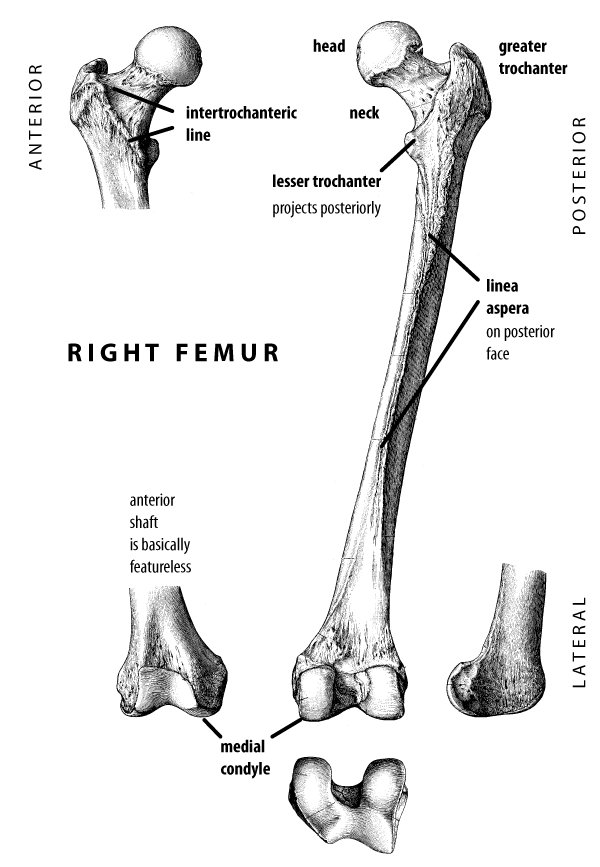
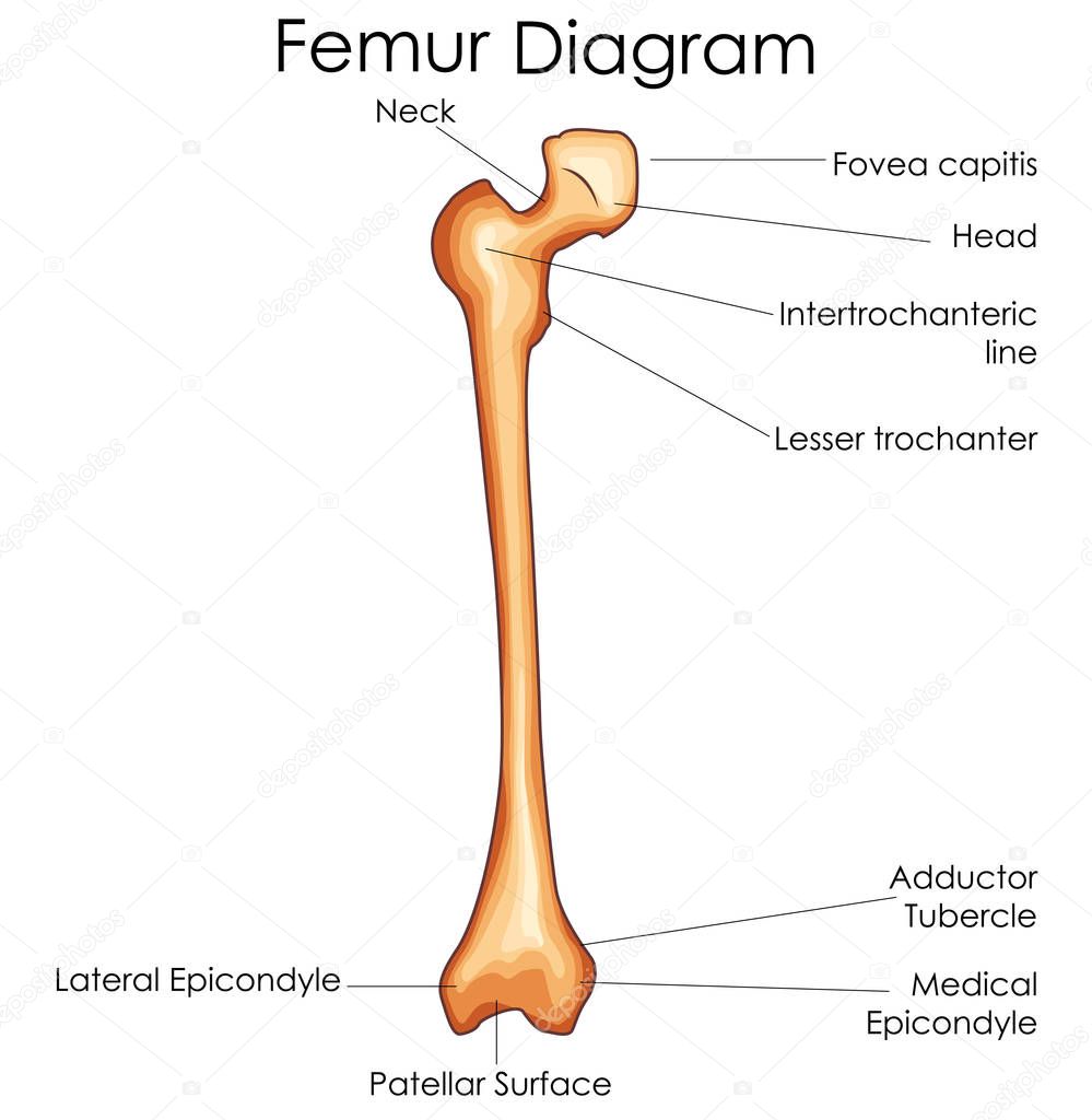
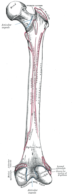
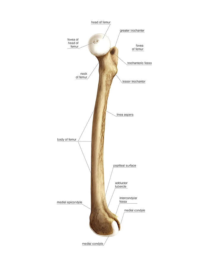
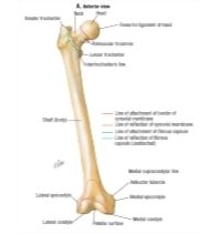
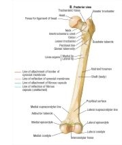
Femur Bone - Anterior and Posterior Markings | GetBodySmart Fovea of Femur Head ( Fovea capitis fe-moris) is a small, pit-like depression on the medial surface of the head, which is also called the fovea capitis. It is as an attachment site for the ligamentum teres ( round ligament; ligament of head of femur ).

Given below is a diagram of the left human hindlimb asseen ...
Femur Labeled Diagram | Quizlet Start studying Femur Labeled. Learn vocabulary, terms, and more with flashcards, games, and other study tools.

Femur bone anatomy: Proximal, distal and shaft | Kenhub The femur bone is the strongest and longest bone in the body, occupying the space of the lower limb, between the hip and knee joints. Femur anatomy is so unique that it makes the bone suitable for supporting the numerous muscular and ligamentous attachments within this region, in addition to maximally extending the limb during ambulation.

Quadratus Femoris - Attachments, Actions & Innervation ...
labeled femur bone - anatomyclass99.z21.web.core.windows.net Femur_labeled anatomycorner.com. femur labeled bones humerus human anatomy skull unlabeled skeleton radius ulna disarticulated anatomycorner main. Femur-Right Anterior View . drawing bones skeleton anatomy bone femur sketch body references printable drawings line thigh human character flash casson anatomi minimal inspiration
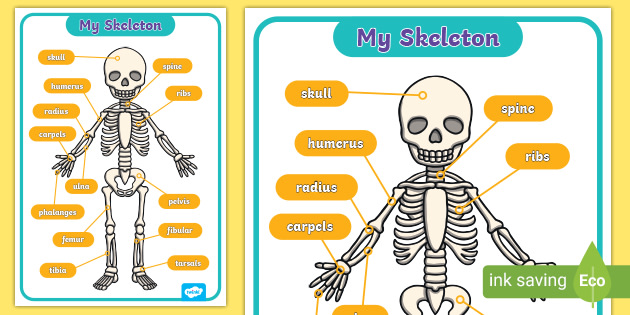
My Skeleton | Human Skeleton Labelled | Display Poster
John Hawks Laboratory

Femoral neck - Wikipedia
REEFERAT REHABILITASI MEDIK PADA POST ROI UNION FRAKTUR SHAFT ...
✓ Medical Education Chart of Biology for Femur Bone Diagram ...
Label the joints - Labelled diagram
Quadrate tubercle - Wikipedia
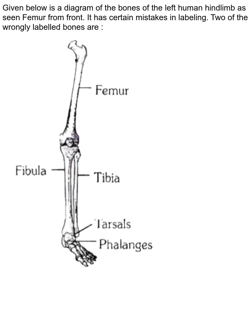
Given below is a diagram of the bones of the left human ...

Left: The initial label configuration for the knee joint. The ...
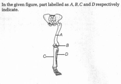
In the given figure, part labelled as A, B, C and D ...

Lateral Healthy Knee - Labelled Medical Illustration ...
Femur Photograph by Asklepios Medical Atlas | Pixels

Femur | KNEEguru
Engraving of the femur hi-res stock photography and images ...
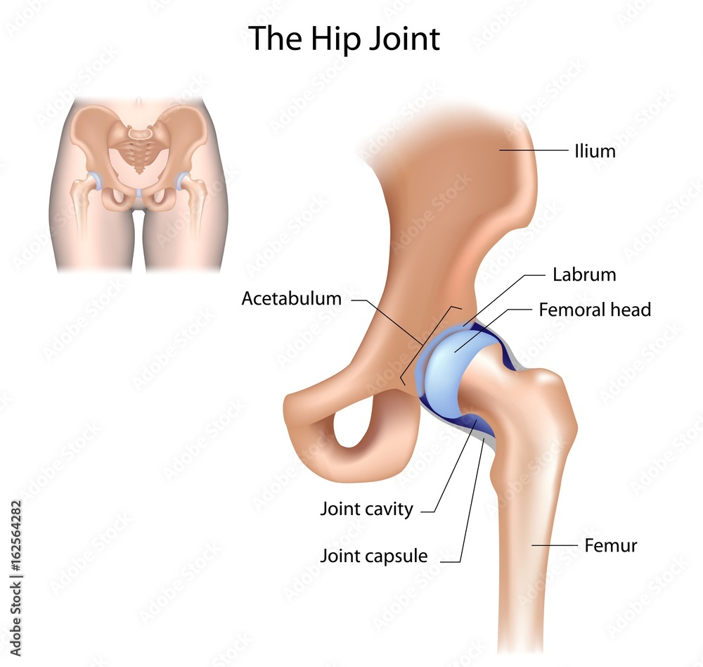
Hip joint structure, labelled Stock Illustration | Adobe Stock

File:Labelled Femur Q Angle.jpg - Wikimedia Commons
The Femur - Proximal - Distal - Shaft - TeachMeAnatomy

Femur, artwork - Stock Image - C020/9159 - Science Photo Library
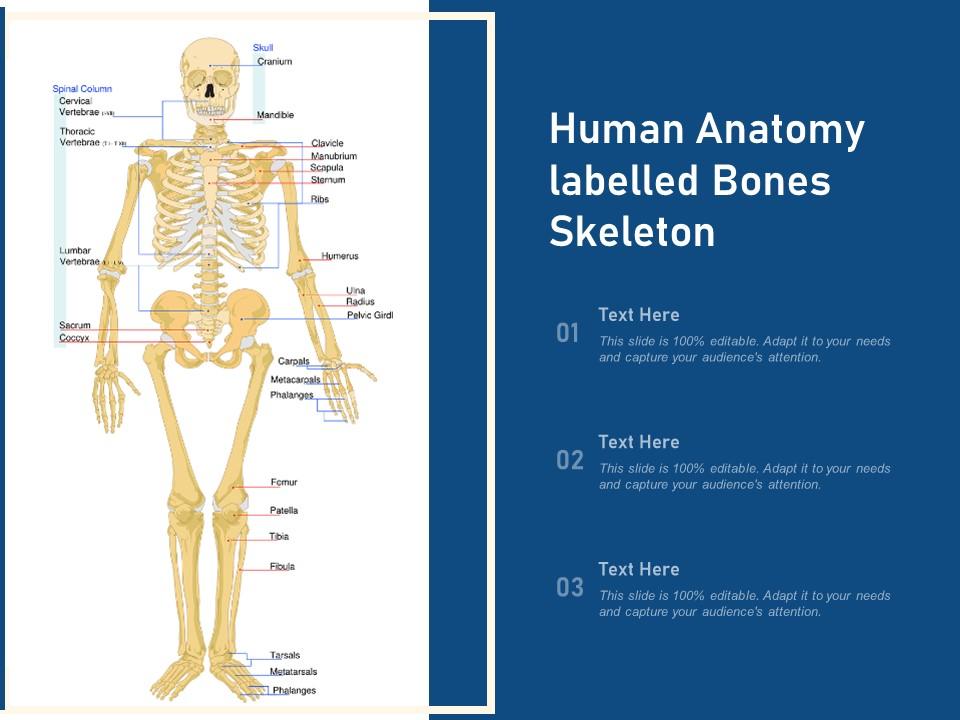
Human Anatomy Labelled Bones Skeleton | Presentation Graphics ...

Femur Bone Anatomy: Labeled Diagram, Quiz, Color-Coded Parts ...

Canine Femur Diagram | Quizlet

Labeling - Right Femur Diagram | Quizlet

Knee Joint (labelled), illustration - Stock Image - C043/4876 ...
4. Bones of the Lower Limb - SimpleMed - Learning Medicine ...

Femur Bone Anatomy: Labeled Diagram, Quiz, Color-Coded Parts ...
Femur: Anterior View
emDOCs.net – Emergency Medicine EducationAn Overview of Femur ...
Femur: Posterior View

Femur | Radiology Reference Article | Radiopaedia.org
Femur

Femur labeled diagram hi-res stock photography and images - Alamy

Consider the diagram given belowParts labelled as A, B, C, D ...

Labelled illustrations of the Palorchestes azael right femur ...

Alila Medical Media | Knee joint labeled drawing. | Medical ...
Post a Comment for "38 labelled femur"