43 neuron diagram with labels
Daisy wild is Best First, label the parts of the neuron on the diagram below. Individual neurons connect one oth… Baca selengkapnya 32+ Neuron Labeling Worksheet Answers Background. 26+ Behaviour Labelling Negotiation PNG. Juni 23, 2021 Posting Komentar The fbi hostage negotiation team developed a five step behavior change. The work in behaviora… Nerve Cells (Neurones) and Synapses Diagram Worksheets NERVE CELLS AND SYNAPSES DIAGRAM WORKSHEET Included in this resource: Nerve Cell Diagram Worksheet - Looking at parts of the nerve cell, students can label and describe the functions (e.g. nucleus, axon, dendrites, myelin sheath etc). Students are also asked to define what a neuron is and the three types or neurone, linked to a simple diagram.
Types of Neurons: Parts, Structure, and Function Interneurons are the most abundant neurons in the body. They act as the signal controllers within the body, relaying important information from one end of the nervous system to the other. The interneurons sit in the middle of other neurons, such as motor or sensory neurons. They are responsible for relaying electrical signals.

Neuron diagram with labels
Identify the parts labeled 1, 2, 3, 4, and 5 of the neuron. ahsan57900 Dendrites, Cell body, Myelin sheath, Schwann cell and Axon are the parts of neuron. The part that is numbered 1 is dendrites which receives signals from other cells and forward it to the next and the part which is numbered 2 is known as cell body that organizes and keeps the cell functional and working. (Get Answer) - Label the parts of the neuron in the diagram. Not all ... Astronomy. ». Label the parts of the neuron in the diagram. Not... Label the parts of the neuron in the diagram. Not all terms will be used. node of Ranvier soma... Label the parts of the neuron in the diagram. Not all terms will be used. node of Ranvier soma Answer Bank axon terminal glial cell axon nerve ganglion dendrite nucleus. Label the structures in the following diagram of a neuron. Draw and label a diagram ...
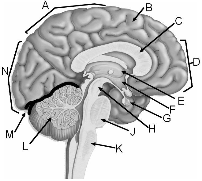
Neuron diagram with labels. Anatomical diagrams of the brain - e-Anatomy - IMAIOS The basic structure of a neuron and an overall diagram of the human nervous system. Meninges : Coronal section . A study of the meninges, ventricles, the circulation of cerebrospinal fluid and an illustration of the dura mater and falx cerebri. ... The user can select to display multiple categories of labels on the illustrations: Cerebral lobes ... Free Nervous System Worksheets and Printables Neuron Printable Clipart and Labeling Sheets - This is an amazing selection of neuron, or nerve cell, clip art for you and your kids to print and label. Inside Out Anatomy: The Brain - This Inside-Out worksheet shows the structure and function of the brain. Kids will color the different parts as they learn about this fascinating body system. Neuron Structure Worksheet Answers - Blogger The diagram below is of a nerve cell or neurone. Sends messages from the cell body to the dendrites of other neurons. Add the following labels to the diagram. Start studying neuron structure worksheet. Students will use evidence from the video to answer questions and gain . What Is a Neuron? Diagrams, Types, Function, and More These neurons allow the brain and spinal cord to communicate with muscles, organs, and glands all over the body. There are two types of motor neurons: lower and upper. Lower motor neurons carry...
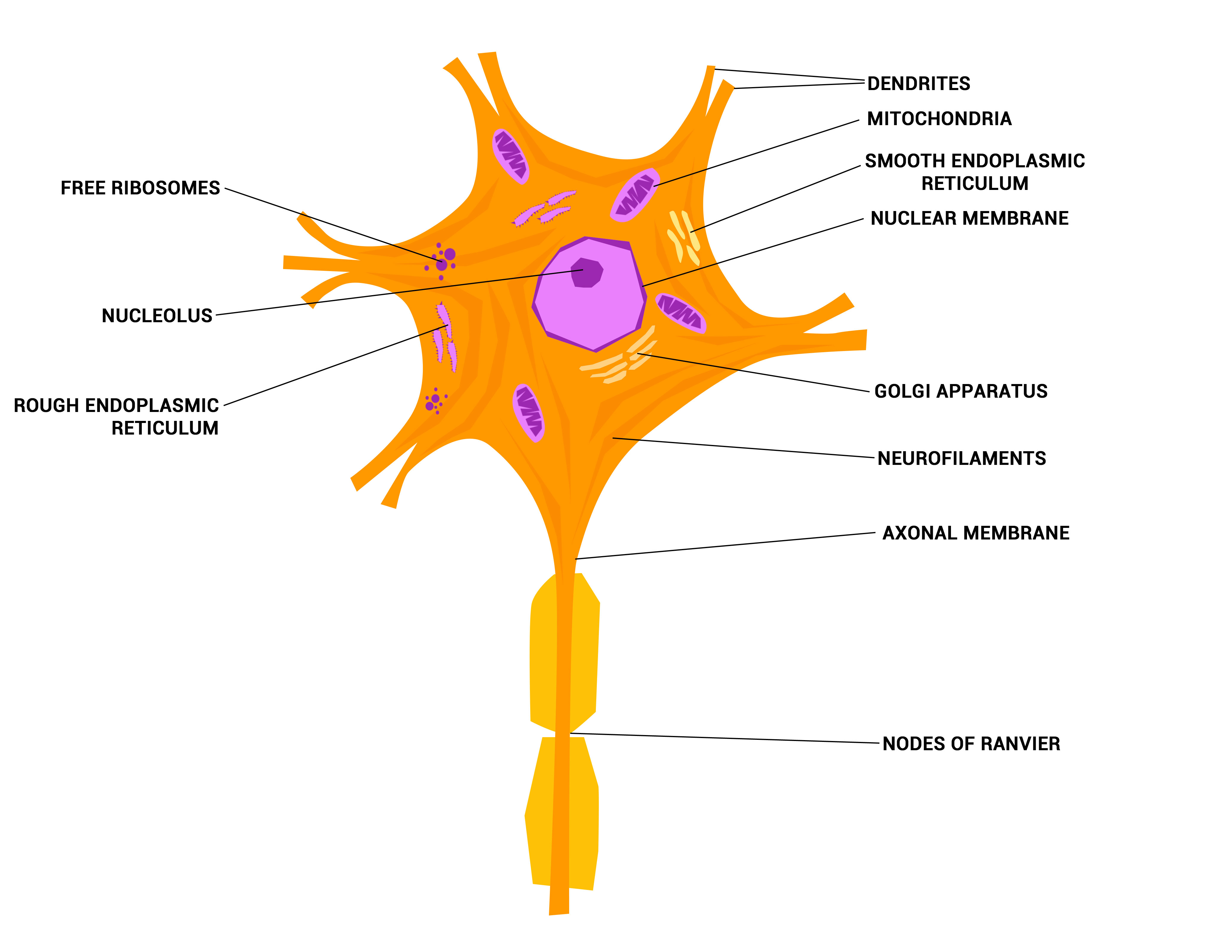
Neuron under Microscope with Labeled Diagram - AnatomyLearner But, first, let's try to identify the following features from a neuron with the help of a labelled diagram. Cell body or perikaryon of a neuron Nucleus, cytoplasm, the plasma membrane of a neuron Nissl bodies in the cell body of a neuron An initial segment of axon and axon hillock Dendrites and axons of a neuron Axolemma and myelin sheath Labeled Neuron Diagram| EdrawMax Template The following labeled diagram shows the parts of a neuron. In order to make it more understandable to the students, we have added all the functions of the Neuron in the labeled diagram. The major parts of the Neuron are Dendrites, Cell Body, Cell Membrane, Axon Hillock, Node of Ranvier, Schwann Cell, Axon Terminal, Myelin Sheath, Axon, and Nucleus. Anatomy of the Brain | Simply Psychology Neurons Neurons are the nerve cells of the central nervous system that transmit information through electrochemical signals throughout the body. Neurons contain a soma, which is a cell body, from which the axon extends. Axons are nerve fibres which are the longest part of the neuron, which conducts electrical impulses away from the soma. Synaptic Cleft | Anatomy, Structure, Diseases & Functions A synaptic cleft is a space that separates two neurons. It forms a junction between two or more neurons and helps nerve impulse pass from one neuron to the other. In this article, we will talk about different aspects of synaptic cleft, its anatomy, and functions. You will completely understand the concept of synapse after reading this article.
Neuron Diagram Labeled | EdrawMax Template It is an effective form of self-assessment, enabling students to check their understanding. In the following diagram, we have illustrated the important parts of the Neuron. In the following Neuron labeled diagram, we have dendrite, cell body, axon, myelin sheath, Schwann cell, a node of Ranvier, axon terminal, and nucleus. Anatomy, Autonomic Nervous System - StatPearls - NCBI Bookshelf Generally, the SNS and PNS motor pathways consist of a two-neuron series: a preganglionic neuron with a cell body in the CNS and a postganglionic neuron with a cell body in the periphery that innervates target tissues. The enteric nervous system (ENS) is an extensive, web-like structure that is capable of function independently of the remainder ... Male Reproductive System: Labeled Diagram of Organs - Study.com Learn the structure and functions of male reproductive anatomy with these labeled diagrams. Updated: 07/14/2021 Table of Contents. Male Reproductive System ... What is a Neuron? Synapses in the Nervous System - Verywell Health Types. In the central nervous system, a synapse is a small gap at the end of a neuron that allows a signal to pass from one neuron to the next. Synapses are found where nerve cells connect with other nerve cells. Synapses are key to the brain's function, especially when it comes to memory. 1 . The term synapse was first introduced in 1897 by ...
Dead Neuron Clip Art at Clker.com - vector clip art online, royalty ...
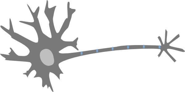
Neurons: Meaning, Types, Functions, Diagrams - Embibe The essential parts of neurons are the dendrite, an axon, cell body, or soma. They can be represented as the branches (dendrite), roots (axon), and trunks (cell body) of a tree (neuron). In a nutshell, for proper coordination among different organs of the human body, neurons play an important role.
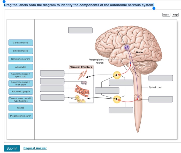
Solved: Drag The Labels Onto The Diagram To Identify The C... | Chegg.com
Histology of neurons: Morphology and types of neurons - Kenhub Neurons have been grouped into two broad categories: those found in the central nervous system (brain and spinal cord) and those in the peripheral nervous system. In the central nervous system, they are found in clusters referred to as nuclei, or in layers also known as laminae. However, in the peripheral nervous system, they are found in ganglia.

Neuron Und Synapse Beschriftetes Diagramm Vektor Abbildung ...
How to determine the number of layers and neurons in the ... - Medium General Structure of Neural Network. A neural network has input layer(s), hidden layer(s), and output layer(s). It can make sense of patterns, noise, and sources of confusion in the data.

Free vector graphic: Science, Neuron, Synapse, Biology - Free Image on ...
Interneurons Function, Diagram & Location | Association Neuron: Example ... A neuron can exist in either the central nervous system (the brain and spinal cord) or the peripheral nervous system (the rest of the body). An interneuron resides within the central nervous ...
7 Best Images of Neuron Label Worksheet - Blank Neuron Cell Diagram ...
A Labelled Diagram Of Neuron with Detailed Explanations Learn more in detail about the Diagram Of Neuron along with their labelling at BYJU'S PreviousNext Diagram Of Neuron A neuron is a specialized cell, primarily involved in transmitting information through electrical and chemical signals. They are found in the brain, spinal cord and the peripheral nerves. A neuron is also known as the nerve cell.

Pin on unlabeled Anatomy
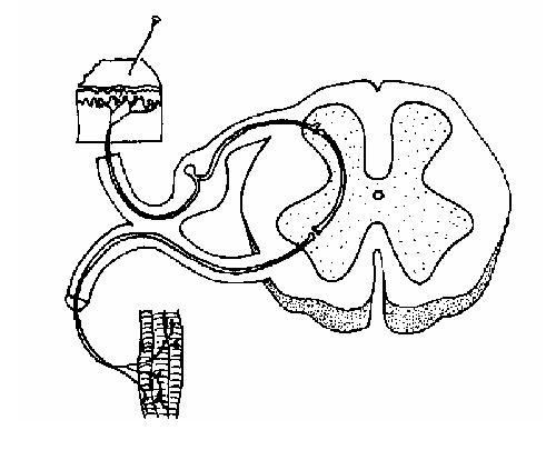
Schwann Cell Anatomy - Human Anatomy - GUWS Medical Figure 25.1 Label this diagram of a motor neuron. Figure 25.2 Label the features of the myelinated nerve fiber. Figure 25.3 Micrograph of a multipolar neuron and neuroglia from a spinal cord smear (100x micrograph enlarged to 600x). -Nerve fiber (axon) general name for processes (either dendrites or axon) of the neuron.

Answer: Ventral Horn (grey matter).
Nervous system: Structure, function and diagram | Kenhub Neurons, or nerve cell, are the main structural and functional units of the nervous system. Every neuron consists of a body (soma) and a number of processes (neurites). The nerve cell body contains the cellular organelles and is where neural impulses ( action potentials) are generated.
Nervous System Worksheet - WikiEducator
drag the labels to their appropriate locations on the diagram of the ... Drag the labels to their appropriate locations on the diagram of the neurons below. use targets of group 1 to indicate the components of neurons. use - 26010782 ... The neurotransmitter is a molecule that travels through the synaptic space forward to the other neuron. Once it reaches the target, ...
Neurons - The crazy wires in our body. | Doc Jana
What are the 12 cranial nerves? Functions and diagram The cranial nerves are a set of twelve nerves that originate in the brain. Each has a different function responsible for sense or movement. The functions of the cranial nerves are sensory, motor ...
Post a Comment for "43 neuron diagram with labels"